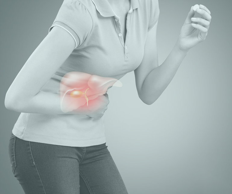
Gallbladder

The gallbladder is a small pouch that sits just under the liver. The gallbladder stores bile produced by the liver. After meals, the gallbladder is empty and flat, like a deflated balloon. Before a meal, the gallbladder may be full of bile and about the size of a small pear.
In response to signals, the gallbladder squeezes stored bile into the small intestine through a series of tubes called ducts. Bile helps digest fats, but the gallbladder itself is not essential. Removing the gallbladder in an otherwise healthy individual typically causes no observable problems with health or digestion yet there may be a small risk of diarrhea and fat malabsorption.
Laparoscopic gallbladder surgery (cholecystectomy) removes the gallbladder and gallstones through several small cuts (incisions) in the abdomen. The surgeon inflates your abdomen with air or carbon dioxide in order to see clearly.
The surgeon inserts a lighted scope attached to a video camera (laparoscope) into one incision near the belly button. The surgeon then uses a video monitor as a guide while inserting surgical instruments into the other incisions to remove your gallbladder.
Before the surgeon removes the gallbladder, you may have a special X-ray procedure called intraoperative cholangiography, which shows the anatomy of the bile ducts.
You will need general anesthesia for this surgery, which usually lasts 2 hours or less.
After surgery, bile flows from the liver (where it is made) through the common bile duct and into the small intestine. Because the gallbladder has been removed, the body can no longer store bile between meals. In most people, this has little or no effect on digestion.