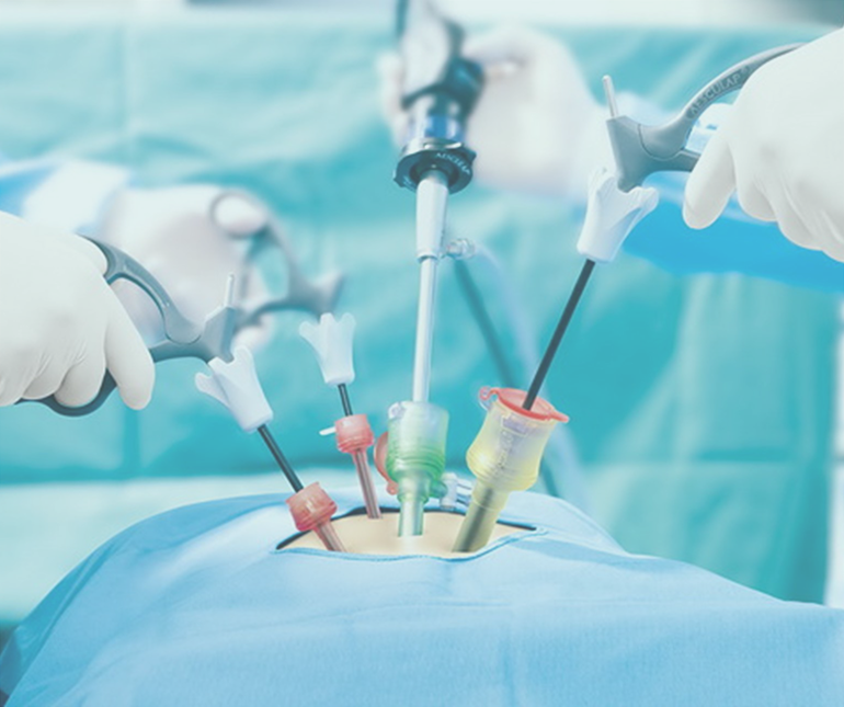
Diagnostic laparoscopy

A procedure wherein a doctor uses a laparoscope to look into the organs and tissues of the abdomen is referred to as a diagnostic laparoscopy. The procedure involves placing a telescope-like instrument through a small incision in the abdomen. The laparoscope is then attached to a high-resolution TV monitor so that proper diagnostic evaluation can be interpreted.
When can Diagnostic Laparoscopy be recommended?
Diagnostic laparoscopy can be very helpful for infertile patients. It becomes easier for the gynecologist to find out the reason for infertility. The doctor can determine whether there are any defects such as endometriosis, ovarian cysts, ectopic pregnancy, tubal disease, genital tuberculosis, fibroid tumors and other abnormalities of the uterus. If any defects are found then they can often be corrected with operative laparoscopy.
Laparoscopy aids in the diagnosis of both acute and chronic abdominal pain. Some of these causes include appendicitis, adhesions or intra-abdominal scar tissue, pelvic infections, endometriosis, abdominal bleeding and, less frequently cancer.
Physicians use laparoscopy to obtain tissue or biopsy to discover the diagnosis of the abdominal mass. The cause of the fluid accumulation (ascites) in the abdominal cavity can be found with laparoscopy.
Why is diagnostic laparoscopy better?
One of the best advantages of laparoscopic diagnosis is the patient can go home a few hours after the surgery. In addition, recovery times are much shorter than when large abdominal incisions are performed. The incisions made are smaller and do not require stitches. The scar also heals early with no noticeable mark. The procedure involves little or no pain and has very few post-op complications.
How is Diagnostic Laparoscopy performed?
Most diagnostic laparoscopy procedures are performed as an outpatient and you can go home the same day the procedure is performed. Prepare yourself prior to the procedure. You should have nothing to eat or drink for six to eight hours before the procedure. Standard blood, urine, or X-ray testing may be required before your operative procedure.
Laparoscopy is done under general anesthesia. After anesthesia, a needle is inserted through the navel, and the abdomen is filled with carbon dioxide gas. As the gas enters the abdomen, it creates a space inside the pelvic area allowing a view of the reproductive organs. The laparoscope is then inserted through the same incision. The laparoscope is connected to the monitor that makes the images larger and easier for the doctor to see. These images are recorded for later viewing and a copy of the same will be given to you on a compact disc.
The following parts can be visible:-
1. Uterus
2. Fallopian tubes
3. Ovaries
4. Bladder
5. Intestines
6. Liver
7. Spleen
8. Appendix
9. Surfaces of the abdominal cavities themselves.
To determine if the fallopian tubes are open a blue solution is injected through the cervix.
If no abnormalities are noted, the instruments are removed and the gas is released. On the contrary, if defects or abnormalities are discovered, the doctor will proceed to operative laparoscopy. After the procedure is done, the doctor may advise a two to three hours stay for anesthesia to wear off.